
Neanderthals probably walked on feet that rolled inward and sat lower to the ground than ours, according to a 3D analysis of fossil heel bones published this month in the Journal of Human Evolution.
Researchers led by Rita Sorrentino of the University of Palermo compared digital models of seven Neanderthal heels with those of 64 Homo sapiens. Neanderthal heels were shorter, wider and taller, and the joints connecting them to the rest of the foot were shaped and angled differently. Three of the bones were digitized with Artec Space Spider, predecessor to the current Artec Spider II ultra-high resolution 3D scanner.
The team says the anatomy reflects bodies built to carry heavy loads over uneven ground. “The robustness of the heel reflects the overall body structure of Neanderthals, which is characterized by a remarkable muscular and bone mass and an extremely intense level of physical activity,” said Maria Giovanna Belcastro, the University of Bologna professor who coordinated the study.

Measuring a Neanderthal heel
The heel bone, or calcaneus, takes the impact of every step and passes force forward as the body moves, so its shape records how its owner walked. The researchers built 3D models from laser, structured-light and CT scans, then used a technique called geometric morphometrics to compare each bone’s overall form as well as specific areas, including the joint surfaces and the ridge where the Achilles tendon attaches.
The modern comparison group included prehistoric hunter-gatherers, Iron Age farmers and foragers, and a postindustrial population buried in Bologna’s Certosa Cemetery.
Flatter feet, heavier loads
The joint shapes point to a foot that habitually pronated, or rolled inward, when bearing weight, and possibly a lower arch along its outer edge. Belcastro said the features “indicate an adaptation aimed at promoting a greater load-bearing capacity on the back of the foot and a high mobility on uneven ground.” The authors call the low-arch theory tentative and say it should be tested with 3D movement modeling.
The human heels varied with lifestyle. Bones from highly mobile groups shared some traits with the Neanderthals, while the city population had lighter heels, which the researchers link to less physical activity and rigid footwear. “The morphology of our skeleton is not only the product of a strict genetic program, but also the consequence of physical stimuli and our daily life habits,” Belcastro said.
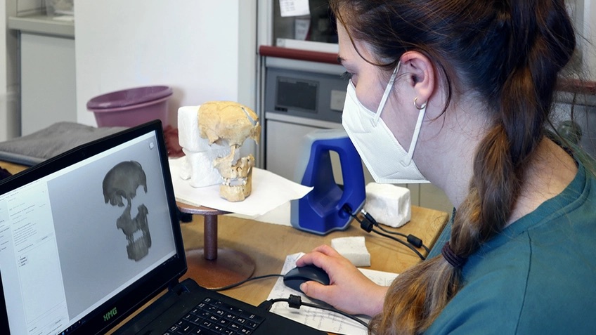
3D scanning for heritage preservation
The authors acknowledge that seven specimens, some partly reconstructed, make a small sample, and that more fossils will be needed to confirm this pattern across the species. Digital models make that kind of expansion easier, since scans can be shared between institutions without moving fragile specimens.
Artec scanners have previously been used to record Homo naledi fossils where they lay in South Africa’s Rising Star cave system, for digitizing hominid finds at Kenya’s Lake Turkana, and to produce 3D replicas of hominid skulls from the Turkana Basin collection, a project paleontologist Louise Leakey described in an earlier 3D Mag interview.
For small bones and teeth, desktop scanners such as the Artec Micro II capture detail at micron-level accuracy, and forensic anthropologists use the same methods to study modern human remains.
[Findings first reported by The Brighter Side of News]