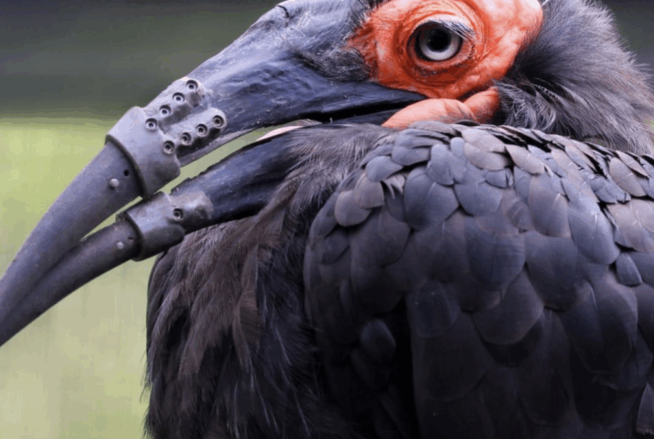

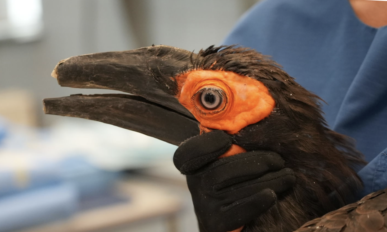
For birds, their beaks almost function like our hands. They gather food, preen feathers and, in many species, help raise their young. When a southern ground hornbill at ARTIS, Amsterdam’s zoo, broke both its upper and lower beak, it was left with two little stumps.
After the accident, it was no longer able to pick food up from the ground or care for its plumage the way a healthy hornbill would. The zoo’s veterinary team responded by partnering with the Faculty of Veterinary Medicine at Utrecht University to design, 3D print, and fit two custom prostheses, one for each half of its beak.
Why did the vets intervene?
ARTIS veterinarian Martine van Zijll Langhout says the decision came down to quality of life. Southern ground hornbills are long-lived, and this one is still young. It had learned to cope, but coping is not the same as thriving. “For me, the most important question is: can an animal exhibit the behavior that belongs to its species?” she said.
Beaks must match the shape of each species’ skull, so a generic replacement was never an option. At Utrecht, the team CT scanned the bird’s head, the remaining stumps and the broken fragments, which keepers had saved. Because CT records internal structure as well as outer form, the specialists could see how the beak sat on the bone and use the fragments to reconstruct its original geometry digitally.
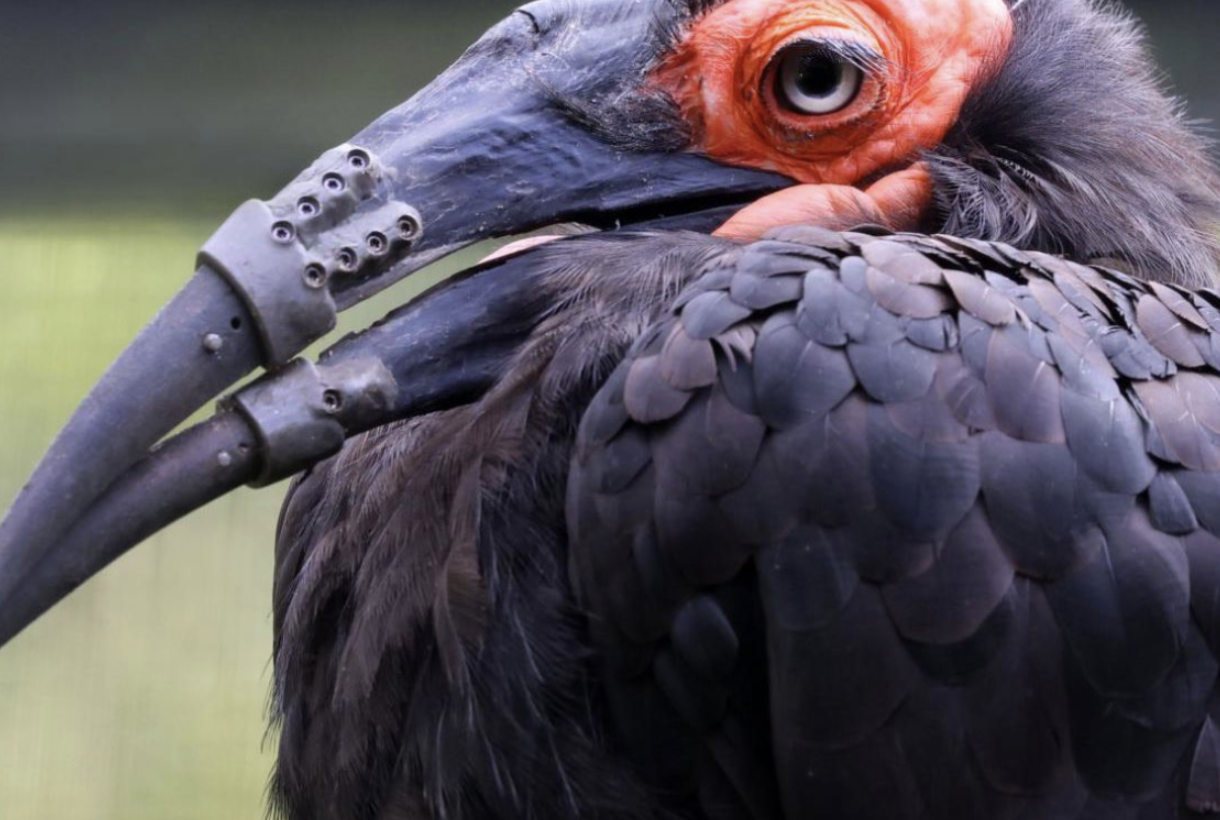
From that model, Joëll Magré of the UMC Utrecht 3DLab designed and printed two plastic beak sections, while Amnovis 3D Lab manufactured the titanium components that anchor them. The result closely follows the shape of the original beak, ensuring a comfortable fit for the bird.
Surgically attaching the beak
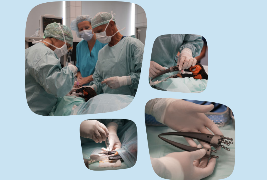
Orthopedic surgeons Bjorn Meij and Lucinda van Stee fixed the prostheses in place with titanium parts and screws, in a project van Zijll Langhout initiated with avian specialist Yvonne van Zeeland. Keepers helped plan the procedure, including what to do if it failed.
The main concern was rejection. Plastic has no sensation, and nobody could be sure the bird would know how to use its new beak. If it stopped eating, the prostheses would come off again, which is why they were screwed rather than permanently fixed. “You should always be able to intervene with such a drastic treatment if it turns out that it is not in the interest of the animal,” van Zijll Langhout said.
That fallback was not needed. The hornbill began testing its new beak the same afternoon, picked a mouse up from the ground the next morning and was eating unaided later that day. Its handling should improve as it gets used to the added weight.
The real test is whether the bird can use the prostheses for life without further surgery. Spare sections have already been made in case one breaks. If the results hold, the team plans to publish its method so that veterinarians elsewhere can learn from it, though van Zijll Langhout cautions that every species’ beak differs in shape and function.
Mr Stubbs proves it’s not a one-off
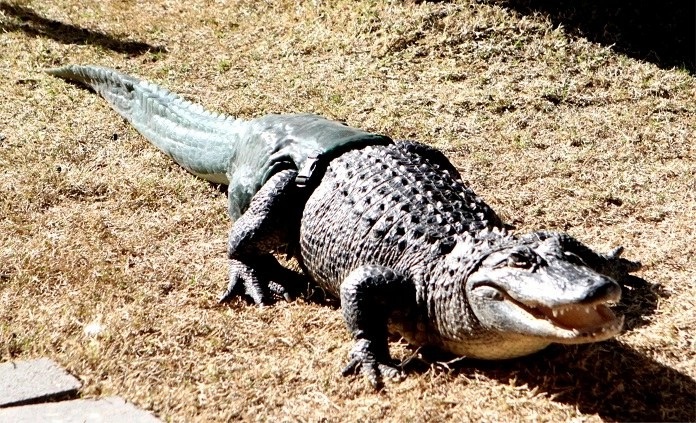
The team’s approach mirrors an earlier case involving a very different animal. Mr. Stubbs, an alligator cared for by the Phoenix Herpetological Society in Arizona, who arrived without a tail after police found him in a truck with 31 other alligators. Without it, he struggled to swim, walked poorly and lost out to faster alligators at feeding time.
Here too, the solution relied on a mixed team: an anatomist from Midwestern University and orthopedic specialists from The CORE Institute who normally treat human patients. And again, the work began with a physical reference. With no tail to measure, Dr. Justin Georgi selected a tail cast from a deceased alligator of similar size.
The capture method differed. Where the hornbill project needed CT to reveal internal anatomy, the tail only required an accurate record of its surface. In this case, the cast was 3D scanned with an Artec Eva handheld structured-light scanner, after an earlier laser scan failed to capture enough detail. The model was refined in Artec Studio, fitted to Mr. Stubbs’ stump and 3D printed as the master for a silicone mold, from which several tails were cast.
Both projects also plan for the long term. ARTIS has spare beak parts ready, while Mr. Stubbs, who could live another 50 to 60 years, may need as many as 40 tails as he grows. In each case, the digital model is the lasting asset, since it can be adjusted and reprinted whenever a replacement is due.
The outcomes match as well. Mr. Stubbs wears his tail without signs of stress and now swims, dives and lunges at food like the alligators around him. For animals whose injuries once meant a lifetime of compromise, scanning and 3D printing have made it practical to restore what was lost, and with it the behavior that depends on it.
Find out about Mr Stubbs’ backstory here. Or discover Artec 3D scanning for healthcare for the company’s other digital healthcare solutions.