
Summary
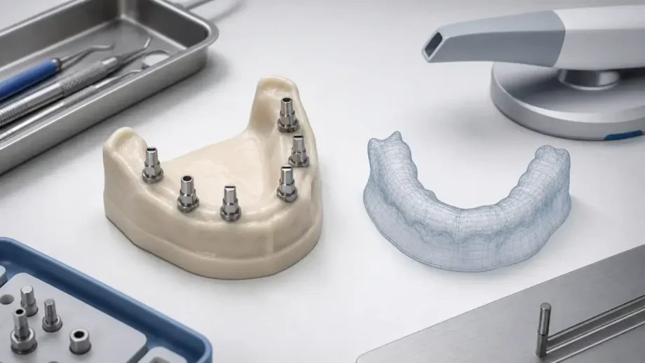
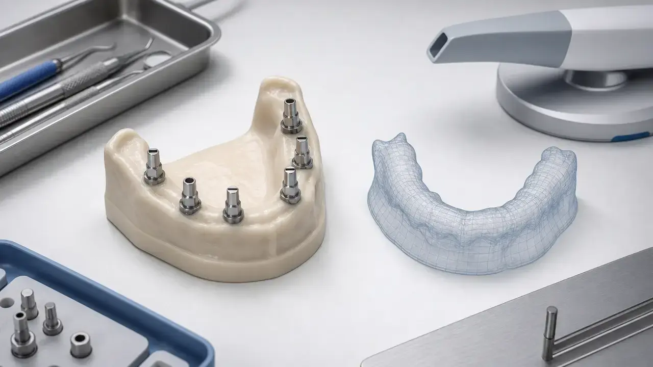
Dental photogrammetry is mainly used to capture implant positions and angulations in full-arch or multi-implant cases. In practice, it often complements rather than replaces intraoral optical scanning, because implant-coordinate capture, soft-tissue recording, and occlusal registration are different measurement tasks with different error models and workflow weak points. [5] [6] [7]
What Dental Photogrammetry Measures — and What It Doesn’t
At-a-glance: measurement task split
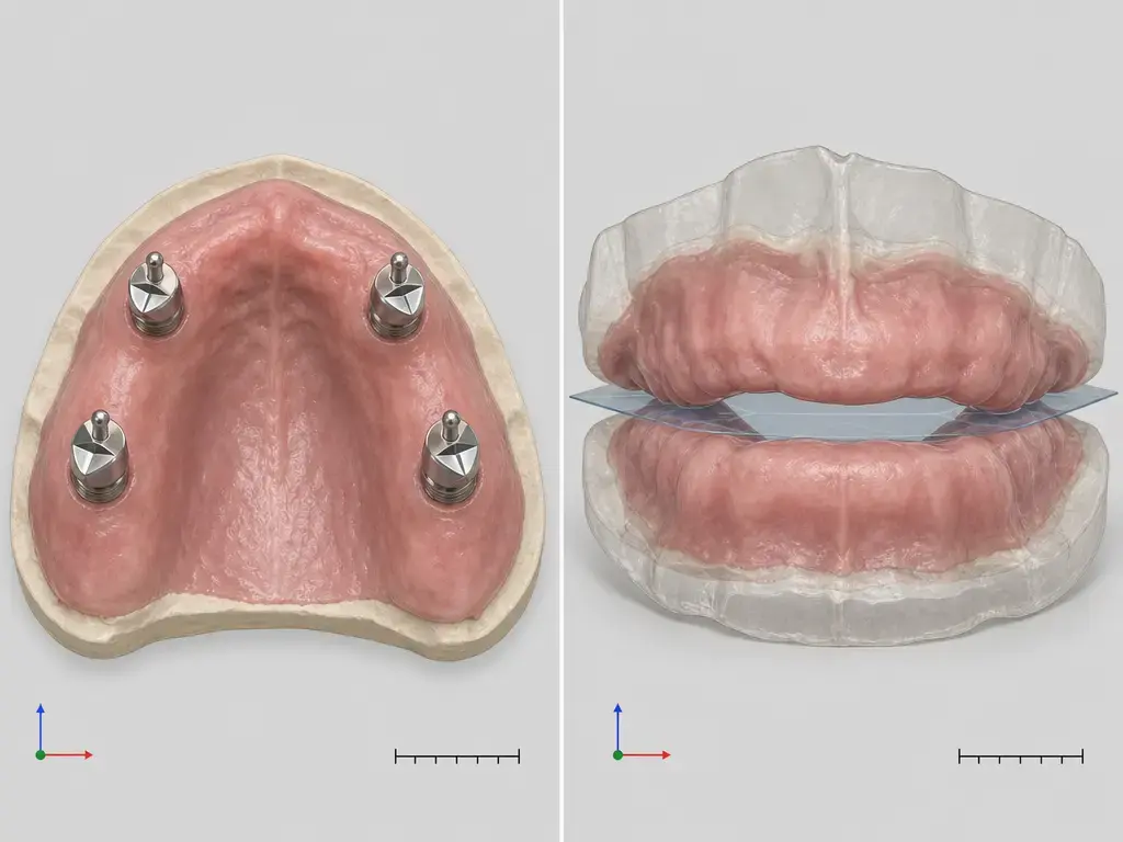
| Implant coordinate capture | Soft-tissue / occlusion surface capture |
|---|---|
| Determines the 3D position and orientation of implant-related markers or scan bodies. | Reconstructs mucosa, teeth, opposing arch surfaces, and bite relationships. |
| Main question: where are the implants, and how are their axes oriented relative to one another? | Main question: what is the shape of the tissues and how do the arches relate in occlusion? |
| Common failure modes: seating error, marker visibility, library mapping, coordinate conversion. | Common failure modes: landmark scarcity, frame stitching, mobile tissue, saliva, and motion. |
These tasks have different error models and are commonly captured with different methods or devices. [1] [5] [6] [7]
In metrology terms, accuracy is better discussed through trueness and precision than as a single floating “micron accuracy” claim. In complete-arch implant work, that distinction matters because one dataset is often used to solve implant coordinates while another is used to record soft tissue and occlusion. [1] [6] [7]
That split becomes more important in edentulous arches. A systematic review on edentulous IOS found that these arches may lack clear anatomic landmarks, which can induce stitching errors, and noted that some authors add landmarks to help stitching. The same review reported higher discrepancies when digitizing mobile or poorly traceable structures such as peripheral borders and soft palate areas. [5]
So when readers ask what dental photogrammetry captures, the practical answer is narrow: primarily implant position and angulation. It does not, by itself, answer every prosthetic question, and it should not be treated as interchangeable with a full soft-tissue and occlusion scan just because both sit inside a digital impression workflow. [5] [6] [7]
Definitions and Device Classes
Photogrammetry vs stereophotogrammetry (SPG) vs intraoral photogrammetry (IPG)
Photogrammetry is the general principle of deriving 3D information from multiple images. In the implant-impression literature summarized here, the strongest comparative evidence is mainly about stereophotogrammetry, or SPG: dedicated systems that record implant targets or scan bodies and calculate their relative 3D coordinates in complete-arch implant cases. That is narrower than saying all photogrammetry workflows share the same evidence base. [6] [8]
In this article, extraoral photogrammetry / SPG refers to workflows in which implant targets are captured by a dedicated camera-based system and then combined with other records as needed. Intraoral photogrammetry, or IPG, is a broader workflow label for handheld or integrated systems that combine implant-coordinate capture with intraoral procedures. That distinction matters because current reviews directly comparing methods are centered on SPG versus IOS, not on every newer integrated or smartphone-based implementation. [6] [7] [8]
That is also why “photogrammetry” should not be used as a synonym for SPG. Some current devices are described by manufacturers as hybrids. Straumann’s 2025 iCam flyer, for example, describes a handheld unit with four cameras and one projector that combines photogrammetric and structured-light scanning techniques. That is a device-architecture description, not independent proof that all such hybrids share the same performance evidence. [16]
Scan body / transfer taxonomy
Component taxonomy matters. Workflows may be implant-level, multi-unit-abutment-level, or based on proprietary markers or transfer geometries, and that choice affects compatibility, reference geometry, and later library mapping in CAD. In the 2025 in vitro comparison used here as a workflow example, the reference geometry was explicitly built around scan bodies and a defined coordinate system, showing how deeply the analysis depends on component choice rather than on scan quality alone. [10]
The same point appears in manufacturer documentation, which describes PIC transfers, ICamBodies, and vendor-specific scan body systems rather than a universal marker standard. That does not make the workflows incomparable, but it does mean an apparently accurate capture can still fail downstream if the wrong component level or library is attached to the dataset. [13] [15] [17]
Why Full-Arch Implant Scanning Is Harder Than Single-Unit Work
Full-arch implant scanning is harder because the measurement span is longer and the anatomy is often less cooperative. In edentulous or nearly edentulous arches, IOS has fewer stable landmarks to follow, so stitching errors can accumulate across the span rather than staying local to one tooth or one short segment. [5] [7]
The tissue side is also less stable than the implant side. Mobile mucosa, saliva, blood, reflective components, partial obstruction of marker dots, and patient movement all affect data capture differently. A scan body that is slightly mis-seated creates one kind of geometric error; a soft-tissue scan distorted by mobility or poor traceability creates another. Treating those as the same problem is one reason complete-arch discussions often become confusing. [5] [10] [13]
A practical way to think about the difficulty is as a stack of recurring failure points across methods. [5] [7]
- scan-body/transfer seating error
- marker visibility/occlusion
- stitching accumulation in IOS
- calibration/verification gaps
- file alignment + implant library mapping errors
- operator + patient factors
Dental Photogrammetry Accuracy — What the Evidence Actually Measures
Metric taxonomy
Before any numbers, the terminology needs to be fixed. ISO 5725-1 treats accuracy through trueness and precision. In this literature, trueness means closeness to a reference, while precision is repeatability across repeated captures. Those are not interchangeable, and neither one is automatically a proxy for final prosthesis fit. [1]
The endpoint matters just as much. Common endpoints include RMS surface deviation from mesh-to-mesh comparison, linear or distance deviation between implant-related reference points, Euclidean 3D deviation where studies explicitly compute a single spatial distance, angular deviation in degrees, and inter-implant distance. RMS in micrometers is not the same thing as distance deviation in micrometers, and neither is the same thing as angular deviation in degrees, so collapsing them into one “accuracy number” is methodologically wrong. [6] [7] [10]
Evidence ladder
The safest reading order is standards and terminology first, then systematic reviews and meta-analyses, then tightly framed primary studies that explain their reference methods, and only after that manufacturer claims. Many disagreements in this topic come less from raw device behavior than from different test objects, reference methods, and reported endpoints. [1] [4] [6] [7]
What systematic reviews/meta-analyses conclude
In a 2025 systematic review and meta-analysis of complete-arch implant impressions, 13 studies met the criteria, including 3 in vivo and 10 in vitro studies. The compared arches ranged from four to eight implants, and SPG reported higher accuracy than IOS in 10 of 13 studies. The included studies used reference methods such as extraoral scanners, industrial optical scanners, coordinate measuring machines, or combined reference-cast-plus-scan workflows. [6]
In that same 2025 review and meta-analysis, for the surface trueness endpoint in in vitro studies referenced by extraoral or industrial scanners and related reference workflows, SPG showed reported ranges of 5.18–48.74 μm, while IOS showed 14.8–67.72 μm. For the surface precision endpoint in in vitro studies using those reference workflows, SPG showed 0.10–5.46 μm, while IOS showed 3.90–37.07 μm. For the angular trueness endpoint in in vitro and in vivo studies referenced against scanner- or cast-based controls, SPG showed 0.24°–0.80°, while IOS showed 0.28°–1.74°. [6]
A later systematic review and meta-analysis, published online in 2025 and assigned to a 2026 journal issue, searched studies from January 2015 to April 2025 and included 14 studies. It evaluated accuracy by distance deviation, angular deviation, and RMS error for both trueness and precision, and its pooled analyses favored photogrammetry over IOS for distance-deviation trueness (P=.001) and angular-deviation trueness (P=.02). The precision analyses also favored photogrammetry for distance deviation (P=.01) and angular deviation (P<.001). [7]
Those findings are directionally consistent, but they do not justify a universal performance figure for “photogrammetry.” The reviewed studies are heterogeneous in reference method, implant number, marker system, arch condition, and endpoint. A workflow that looks excellent by RMS against a lab-scanner reference may not map one-to-one onto a study reporting Euclidean implant-position deviation against a CMM or industrial optical scanner. [6] [7] [10]
System-specific review data reinforce that point. A 2023 systematic review of stereophotogrammetry systems reported that, among the commercially available PG systems discussed in the review, only PIC and iCam4D had been independently analyzed in the reviewed studies. In those datasets, PIC showed trueness of 10–49 μm and precision of 5–65 μm, while iCam4D showed trueness of 24–77 μm and precision of 2–203 μm. These are review-level ranges tied to particular study designs, not a brand ranking or a promise of chairside performance in every clinic. [8]
Evidence map
| Source | Evidence level | Method class | Endpoint(s) | Takeaway |
|---|---|---|---|---|
| Pozzi 2025 [6] | Systematic review + meta-analysis | SPG vs IOS | Surface, linear, angular, inter-implant | SPG was favored in most included studies, but study design and reference methods varied. |
| Altalla 2025/2026 [7] | Systematic review + meta-analysis | PG vs IOS | Distance, angular, RMS | Pooled results favored PG for distance and angular trueness and precision. |
| Gómez-Polo 2023 [8] | Systematic review | SPG systems | Trueness, precision, scan time, satisfaction | Review-level ranges were system-specific, and only some commercial systems had peer-reviewed evidence. |
| Abuduwaili 2025 [10] | In vitro | Conventional vs IOS vs two PG systems | X/Y/Z, 3D, angular, RMS | A defined reference and coordinate system are essential for interpreting reported discrepancies. |
| He 2026 [9] | Systematic review | Multiunit implant prostheses | Misfit thresholds | Proposed thresholds exist, but no consensus defines one settled acceptable misfit for complete arches. |
Study examples
The 2025 in vitro study by Abuduwaili and colleagues is useful because it shows how numbers are constructed. Its reference was a laboratory scanner with reported precision of 5 μm, exported as STL. The investigators then created a coordinate system on the reference scan bodies, setting the origin on scan body A, defining axes, and calculating x/y/z, 3D Euclidean-type deviation, angular discrepancy, and RMS. That design is a reminder that an accuracy result is always a relationship among a test scan, a reference, and a chosen endpoint. [10]
As historical context, a 2013 in vitro study had already compared a photogrammetric complete-arch impression technique with a conventional one by using a coordinate measuring machine as the control/reference. It is useful mainly as an early peer-reviewed example of implant-position recording against a metrology-style reference, not as a direct benchmark for current device generations. [11]
Passive fit: what can be said responsibly
Claims about passive fit need the most restraint. A 2026 systematic review on misfit in multiunit implant-supported fixed prostheses reported proposed thresholds from 10 to 150 μm, but also concluded that no consensus exists on what constitutes clinically acceptable misfit, especially for multiunit complete arches. So a single μm or angular number should not be presented as settled clinical law, and neither SPG nor IOS accuracy data should be over-read as direct proof of final clinical fit without fabrication and verification context. [9]
Workflow: From Scan Bodies to CAD/CAM
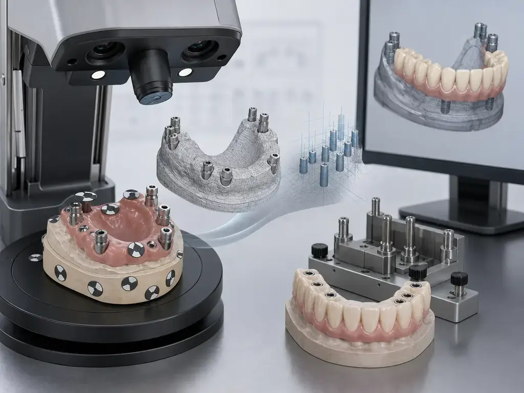
A practical dental photogrammetry workflow separates implant-coordinate capture from surface capture, then reconnects them in software. The point is not to create extra files for their own sake, but to avoid asking one dataset to solve two different metrology problems at once. [6] [10] [15]
Step-by-step clinical + lab workflow
- choose components at the implant or MUA level and confirm the matching libraries
- seat and verify scan bodies or transfers
- capture the PG/SPG dataset for implant coordinates
- capture the soft-tissue, arch, and occlusion dataset, often with IOS
- merge the datasets using the software’s supported alignment and library workflow
- complete CAD design and decide the provisional/final strategy
- perform verification before final delivery, whether digitally, physically, or both. [10] [13] [15] [17]
In published and manufacturer-described workflows, the lab may receive a surface mesh, often STL in the published in vitro example, plus an implant-coordinate solution or a proprietary photogrammetry dataset. Some ecosystems then reconstruct implant platforms through library-based mapping rather than from raw surface alone. Exact file types beyond that vary by system, and documentation may be proprietary. Imetric, for example, describes a merged workflow in which a photogrammetry file for implants is combined with an intraoral scan file for soft tissue. [10] [15]
That handoff is where many avoidable errors occur. A correct-looking mesh can be paired with the wrong component library, a valid coordinate file can be transformed incorrectly at merge, and a well-captured implant dataset can be undermined if the wrong level—implant versus MUA—was assumed later in CAD. The workflow is a chain, not a single capture event. [10]
Short error-source split for the handoff stage:
- component/seating: incomplete seating, wrong transfer selection, rotation during capture
- acquisition/environment: reflections, obstruction of dots or markers, saliva, blood, motion, poor angle variation
- digital alignment/library mapping: wrong library, merge drift, coordinate-transform error, CAD assumptions. [10] [13] [15]
Manufacturer guidance helps explain why capture discipline matters. PIC support documentation says a capture typically takes about 1–2 minutes, states that transfer placement accounts for 80% of success, recommends 10–30 degrees of angulation between transfers, suggests off-center views around 30–45 degrees from center, notes that progress may stay at 0% for the first 3–5 photos, and recommends at least 60 megabits per second upload speed. These are workflow claims, not independent comparative evidence, but they illustrate why the process is sensitive to component placement, visibility, and data transfer conditions. [13]
Extraoral Photogrammetry vs Intraoral Photogrammetry vs IOS-Only vs Conventional
The most useful comparison is not which method is best, but which task each one is solving and where each tends to fail. The direct independent evidence base is still strongest for extraoral SPG versus IOS in complete-arch implant impressions, while integrated IPG and smartphone-based approaches are also market realities that should not automatically be treated as evidence-equivalent. [6] [7]
| Method class | Captures | Strengths | Limitations |
|---|---|---|---|
| Extraoral photogrammetry / SPG | Implant markers or scan bodies | Strongest independent comparative evidence for implant-coordinate capture in complete arches | Usually needs separate soft-tissue/occlusion capture and later merging |
| Intraoral photogrammetry (IPG class) | Implant coordinates within a handheld/integrated workflow | May simplify chairside sequence and reduce system switching | Evidence maturity is more product-specific; not all systems share the same review base |
| IOS-only implant scan | Surface plus scan bodies in one digital pass | One-device surface workflow and direct arch/occlusion capture | Long spans, stitching, landmark scarcity, and mobile tissues can degrade results |
| Conventional open-tray/reference workflow | Physical impression and cast-based transfer | Familiar reference workflow and no digital merge at capture stage | Material, cast, and later digitization steps can each add error |
ISO boundaries help keep those classes separate. ISO 20896-1 applies to handheld intraoral surface capture, ISO 12836 applies to mounted digitizers, and ISO 10360-13 is best read here as a metrology analogy about defined test objects, references, and measurement volume rather than as a dental-photogrammetry validation standard. A fair comparison therefore depends on exactly what was captured, how it was referenced, and what still had to be added afterward. [2] [3] [4]
Metrics and Standards — How to Read “Accuracy” Claims
When reading an accuracy claim, ask five questions: What endpoint was measured? Was the evidence in vitro, in vivo, or just a manufacturer page? What reference method was used? Over what span or measurement volume? And is the device solving implant coordinates, surface capture, or both? ISO 5725-1 is the terminology foundation for trueness and precision, but those words are only meaningful once the endpoint and reference are specified. [1] [4]
The standards are boundary markers, not a universal badge. ISO 20896-1 specifies methods for assessing the accuracy of a 3D numerical description of intraoral surfaces acquired directly from a patient with a handheld scanning device, and ISO lists that 2019 edition as confirmed in 2025 while also showing ISO/CD 20896-1 under development. ISO 12836 applies to mounted digitizers, not handheld scanners. ISO 10360-13 addresses acceptance and reverification tests for optical 3D coordinate measuring systems measuring lengths stated by the manufacturer. Longer spans or larger measurement volumes generally make cumulative error more important, which is why complete-arch claims deserve more scrutiny than single-unit ones. [2] [3] [4]
Dental Applications and Lab Handoff
The clearest application zone is complete-arch implant prosthodontics: full-arch definitive frameworks, immediate-load provisionals, and verification-oriented workflows in which the relative position of multiple implants matters more than any single local surface patch. That is also the zone where the review evidence comparing PG/SPG with IOS is most developed. [6] [7]
For the lab, accurate implant coordinates matter only if the handoff stays coherent. The component level has to match the library, the library has to match the captured geometry, and the coordinate solution has to merge correctly with the soft-tissue and occlusal record. The 2025 in vitro study is helpful here because it explicitly used reference scan bodies, coordinate construction, and conversion steps, showing why wrong library or component mapping can compromise a case even when the underlying capture was good. [10]
Limitations and Failure Modes
Photogrammetry or SPG is not the entire prosthetic record. It is a strong way to capture implant coordinates, but it does not automatically solve emergence profile, movable mucosa, opposing arch form, or bite by itself. A good implant-coordinate dataset can still produce a poor outcome if the soft-tissue record, merge, design assumptions, fabrication, or verification step adds error later. [5] [9]
A second limit is practical sensitivity during capture. Manufacturer guidance for smartphone-based capture emphasizes transfer placement, angle variation, reflections, focus, and upload conditions, which indirectly shows that even a strong measurement method is still vulnerable to ordinary chairside mistakes. Meanwhile, the edentulous IOS literature shows that mobile and poorly traceable tissues remain problematic on the surface-capture side of the workflow. [5] [13]
A third limit is interpretive. Because clinically acceptable misfit thresholds are still unsettled for multiunit complete arches, it is a mistake to treat any single trueness or precision figure as a complete clinical guarantee. Accuracy data are useful, but they do not eliminate the need for verification before final delivery. [9]
-
Component & hardware
-
incomplete seating of scan bodies or transfers
-
wrong component level or wrong marker set
-
Acquisition & environment
-
marker obstruction or poor line of sight
-
saliva, blood, shadows, and reflections
-
motion from operator or patient
-
Digital & downstream
-
merge or alignment drift between implant and surface datasets
-
coordinate-transform mistakes
-
wrong implant library or platform mapping
-
CAD assumptions that are never independently verified. [10] [15] [9]
Current Research and Market Context
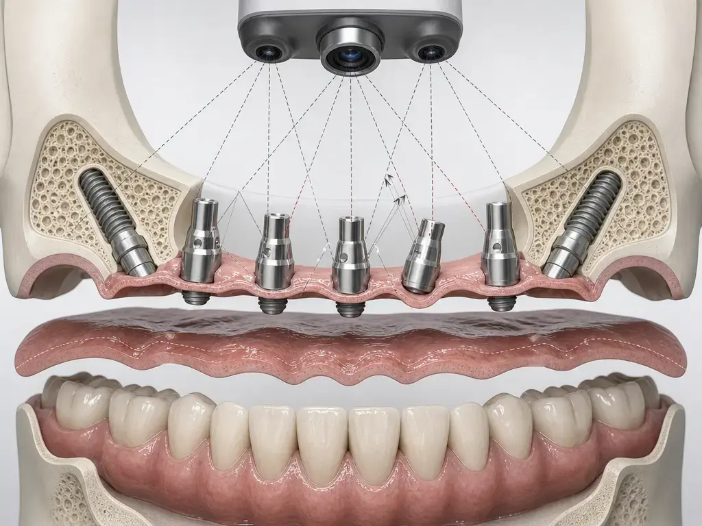
The market is moving from stand-alone SPG camera systems toward integrated and faster workflows. PIC positions its PIC app as smartphone-based photogrammetry and claims implant positions and angulations can be captured in about 1–3 minutes. SHINING 3D positions Aoralscan Elite as a combined IOS plus intraoral photogrammetry device and lists “Precision for IPG 5 μm” on its product page. Imetric describes ICam as a four-camera system with sub-5-micron precision, while Straumann’s 2025 flyer describes iCam as a handheld unit with four cameras and one projector combining structured light and photogrammetry. ClaroNav’s MicronMapper page claims an average scan time of 10 seconds and “No Warm Up. No Pre-Calibration. No Delay.” These are manufacturer claims, not independent comparative proof. [12] [14] [15] [16] [17]
The research message is more conservative than the market message. The strongest independent comparative evidence still centers on SPG versus IOS for complete-arch implant impressions, and the newer meta-analyses do favor photogrammetry in key distance and angular endpoints. But that does not mean every integrated IPG workflow or smartphone-based workflow has the same evidence maturity today. It is worth separating workflow innovation from independently replicated accuracy evidence. [6] [7] [8]
What Dental Photogrammetry Can — and Cannot — Solve
Dental photogrammetry can solve implant-coordinate capture very well in the specific context where the evidence is strongest: complete-arch or multi-implant digital impressions. What it cannot do, by itself, is guarantee final prosthesis fit or replace every other record in the case. Clinically relevant fit still depends on the full chain—component seating, acquisition, alignment or library mapping, fabrication, and verification—and current literature still does not support one universally settled misfit threshold for complete arches. [6] [9]
FAQ
What is dental photogrammetry in implant dentistry?
Dental photogrammetry is a way of recording the 3D position and angulation of implants by analyzing images of implant markers or scan bodies rather than relying only on stitched surface images. In implant dentistry, its main use is complete-arch or multi-implant coordinate capture, often alongside a separate surface scan for soft tissue and occlusion. The best comparative review evidence for this use case is mainly about SPG systems in full-arch implant impressions rather than every product marketed under the broader photogrammetry label. (systematic review/meta-analysis) [6] [8]
What is intraoral photogrammetry, and how is it different from stereophotogrammetry?
Intraoral photogrammetry usually refers to handheld or integrated workflows that capture implant coordinates within an intraoral scanning-style process. Stereophotogrammetry, as discussed in the main comparative reviews, more specifically refers to dedicated implant-position capture systems used in complete-arch cases. The practical difference is not just device shape but evidence maturity: the strongest review data directly compare SPG with IOS, while integrated IPG workflows are more product-specific and often supported partly by manufacturer documentation. (systematic review/meta-analysis; manufacturer claim) [6] [7] [14] [16]
How accurate is dental photogrammetry for full-arch implants?
Accuracy has to be answered by endpoint, study type, and reference method. In a 2025 systematic review/meta-analysis of complete-arch implant impressions using references such as extraoral scanners, industrial scanners, CMMs, or cast-plus-scan workflows, SPG showed surface trueness of 5.18–48.74 μm, surface precision of 0.10–5.46 μm, and angular trueness of 0.24°–0.80°, while IOS showed 14.8–67.72 μm, 3.90–37.07 μm, and 0.28°–1.74° for those endpoints. A later meta-analysis also favored PG for distance and angular trueness and precision. These are review-level ranges, not a universal chairside guarantee. (systematic review/meta-analysis) [6] [7]
Does dental photogrammetry capture soft tissue and bite by itself?
Often no. Dental photogrammetry usually captures implant coordinates, not the full soft-tissue and occlusal picture by itself. That is why many workflows add an IOS or another surface-capture step for mucosa, opposing arch, and bite. Manufacturer documentation for some systems explicitly describes merging an implant photogrammetry file with a separate intraoral scan file for soft tissue, which matches the general workflow logic seen in the literature. (systematic review/meta-analysis; manufacturer claim) [6] [10] [15]
What is a typical dental photogrammetry workflow from clinic to lab?
A typical workflow is to choose the component level and libraries, seat and verify the scan bodies or transfers, capture implant coordinates, capture soft tissue and occlusion, merge the datasets, design the restoration, and verify before final delivery. The handoff may include a surface mesh plus an implant-coordinate solution or proprietary dataset, and errors can enter at component seating, during acquisition, or during library mapping and alignment. Manufacturer guides add practical variables such as angle variation, visibility, and network dependence. (in vitro; manufacturer claim) [10] [13] [15]
What reference methods do studies use, and why does that choice affect reported trueness and precision?
Reference methods vary, and that is a major reason published values are not directly interchangeable. Some studies use a laboratory scanner with stated precision and then compute x/y/z, 3D, angular, and RMS discrepancies from a defined coordinate system on scan bodies. Earlier work used a coordinate measuring machine as the control/reference. Because trueness and precision are always relative to a reference and an endpoint, changing the reference method changes what the reported number really means. (in vitro) [10] [11]
Sources
Standards
- ISO 5725-1:2023 — Accuracy (trueness and precision) of measurement methods and results — Part 1. https://www.iso.org/standard/69418.html
- ISO 20896-1:2019 — Dentistry—Digital impression devices—Part 1. https://www.iso.org/standard/69402.html
- ISO 12836:2015 — Dentistry—Digitizing devices for CAD/CAM systems for indirect dental restorations. https://www.iso.org/standard/68414.html
- ISO 10360-13:2021 — Acceptance/reverification tests for coordinate measuring systems — Optical 3D CMS. https://www.iso.org/standard/74957.html
Systematic reviews/meta-analyses
- AlHelou et al. (Dent. J. 2023) — Accuracy of intraoral scanner for recording completely edentulous arches (systematic review, PDF). https://mdpi-res.com/d_attachment/dentistry/dentistry-11-00241/article_deploy/dentistry-11-00241.pdf?version=1697607935
- Pozzi et al. (2025) — Photogrammetry versus intraoral scanning in complete-arch digital implant impression (systematic review + meta-analysis; PMCID page). https://pmc.ncbi.nlm.nih.gov/articles/PMC12144927/
- Altalla et al. (J Prosthet Dent, Epub 2025 / issue 2026) — Comparative accuracy of photogrammetry and intraoral scanners in recordings for complete arch implant-supported prostheses (systematic review + meta-analysis). https://pubmed.ncbi.nlm.nih.gov/41241558/
- Gómez-Polo et al. (J Prosthodontics, 2023) — Accuracy, scanning time, and patient satisfaction of stereophotogrammetry systems for acquiring 3D dental implant positions (systematic review). https://onlinelibrary.wiley.com/doi/full/10.1111/jopr.13751
- He et al. (J Prosthodontics, 2026) — Clinical implications of misfit in multiunit implant-supported fixed dental prostheses (systematic review). https://onlinelibrary.wiley.com/doi/full/10.1111/jopr.70231
Primary studies
- Abuduwaili et al. (BMC Oral Health, 2025) — Comparison of photogrammetric imaging, intraoral scanning and conventional impression accuracy of full-arch dental implant rehabilitation: an in vitro study (PDF). https://bmcoralhealth.biomedcentral.com/counter/pdf/10.1186/s12903-025-06029-8.pdf
- Bergin et al. (2013) — An in vitro comparison of photogrammetric and conventional complete-arch implant impression techniques (PubMed record). https://pubmed.ncbi.nlm.nih.gov/24079558/
Manufacturer/official docs
- PIC Dental — PIC app & PIC cloud (product page). https://www.picdental.com/pic-app
- PIC Dental Support — How to capture implant positions with PIC app. https://support.picdental.com/help-center/how-to-capture-implant-positions-with-pic-app
- SHINING 3D Dental — Aoralscan Elite product page. https://www.shining3ddental.com/solution/aoralscan-elite/
- Imetric — Dental Photogrammetry / ICam page. https://www.imetric.com/index.php?id=5
- Straumann — iCam Sales Flyer (PDF). https://www.straumann.com/content/dam/media-center/straumann/en-us/documents/letter/USLIT.1778-Straumann-iCAM-Sales-Flyer.pdf
- ClaroNav — MicronMapper product page. https://www.claronavdental.com/micronmapper
Industry
- Inside Dentistry (2023) — The advantages of photogrammetry in implant dentistry. https://insidedentistry.net/2023/06/the-advantages-of-photogrammetry-in-implant-dentistry