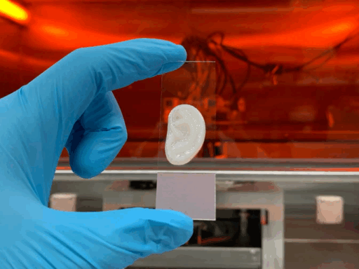

A revolutionary study from Yokohama National University has presented an ingenuous way to create multi-directionally oriented collagen tissues using 3D printing and fluidic technology. This novel approach is a safer, more precise alternative to existing methods and could open new frontiers in biomedical engineering, particularly in tissue modeling and regenerative medicine.
Collagen, a vital structural protein in the human body, plays a critical role in tissue stability and mechanical strength. Despite its prevalence, the orientation of collagen fibers, which is essential for tissue function, is notoriously difficult to replicate in lab-grown artificial models. Conventional techniques like magnetic alignment and electrospinning have their limitations, including residual magnetic beads, the use of volatile organic solvents, or lack of control over direction.
In the study, recently published in ACS Biomaterials Science and Engineering, the research team introduced an innovative 3D-printed fluidic system. “By utilizing a technique that uses flow to orient collagen fibers and cells, it is possible to fabricate complex oriented tissues with multiple directions in flow channels constructed using a 3D printer,” Kazutoshi Iijima, associate professor and co-author of the study, said in a press-release. This breakthrough allows scientists to recreate the complex, layered architecture seen in tissues such as the dermis and skull bone, where fibers naturally run in both horizontal and vertical directions.
“This system will lead to the customization of tissue-specific models using fine, multidirectionally oriented biomaterial scaffolds for the preparation of various oriented biological tissues,” added Professor Shoji Maruo.
With its blend of precision 3D printing and biological compatibility, this innovation marks a stride toward the future of regenerative medicine and advanced biomaterials. The researchers now aim to refine this technique for applications in transplantation and in vitro tissue modeling.